"Attention Deficit Hyperactivity Disorder is characterized by persistent Inattention and/or Hyperactivity - impulsivity which interferes with normal development and functioning" (Diagnostic and Statistical Module of Mental Disorders-5). Inattention may manifest as wandering off task, inability to sustain focus, and disorganized behavior which is not due to defiance on the part of the child. Hyperactivity refers to excessive movements such as fidgeting, tapping or even excessive running. Impulsivity refers to actions which are carried out by the child without any forethought or consideration. ADHD has been classified under Intellectual Disabilities in DSM-V (Diagnostic and Statistical Module of Mental Disorders).
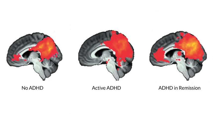
Image 1.3 shows the difference in brain signals in three conditions.
Image source - Aaron T. Mattfeld and others, Brain differences between persistent and remitted attention deficit hyperactivity disorder, Brain, Volume 137, Issue 9, September 2014, Pages 2423–2428, https://doi.org/10.1093/brain/awu137
Neural development is also seen to be slower in them meaning that brain maturation is delayed (Shaw et al, 2012 and Shaw et al, 2007). This leads to delay in achieving developmental milestones as well as cognitive deficits. A meta-analysis study conducted in 2005 described executive function deficits with moderate effect sizes in children with ADHD including response inhibition, vigilance, working memory, and planning (Willcutt et al, 2005). It has been seen that almost every neuropsychological aspect is significantly impaired in ADHD though the correlation coefficients were moderately positive (small effect sizes) (Drechsler et al, 2020).
It was earlier believed that ADHD was caused due to a defect of inhibitory self-control (Barkley et al, 2005). This concept was logical since the most prominent feature seen in children with ADHD was hyperactivity which leads to the conclusion that the child is unable to maintain self control. Another model which looked to describe the pathophysiology of ADHD described two pathways in ADHD namely, Inhibition/Executive function pathway and the Motivation/Aversion pathway (Castellanos et al, 2006 and Sonuga-Barke et al, 2002).
Later, other pathways were also included such as time processing but it is difficult to point out the exact number and type of pathways involved in ADHD since the behavioral manifestations are varied which in turn, are controlled by a network of several pathways (Sonuga-Barke et al, 2010). The important pathways involved have been mentioned in the text.
The neural pathways do not connect at the same rate making it difficult to focus and stay attentive. Studies have described an essential resting state network, known as the Default Mode Network (DMN), which shows high activity when a person is awake and resting and its activity levels decrease as we increase our attention. Along with this there is another network, known as the cognitive control network which shows increasing activity with rising attention levels. We can clearly see that there is an inverse relation between the Default Mode Network and the Cognitive Control Network which is either diminished or absent in children with ADHD and this might be the cause of attention lapses seen in ADHD (Posner et al, 2014)
Meta-analysis studies have also shown that there are consistent changes in brain activation patterns in ADHD seen as hypoactivation of the frontoparietal network during executive functions and hypoactivation of the ventral attention system during activities which require attention (Cortese et al, 2012).
Alongside, there is defective reward processing and Motivational processes. This is an important feature since most of the models which describe ADHD provide altered sensitivity to reward as an important factor in the causation of ADHD (Sonuga-Barke et al, 2011 and Tripp et al, 2008).
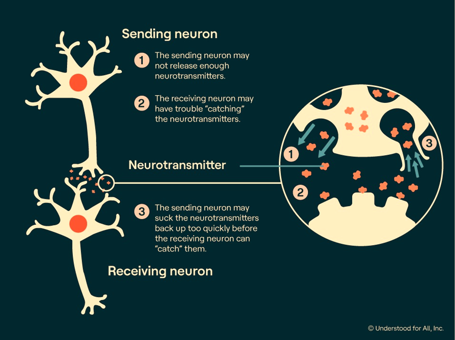
Image 1.4 depicts the defects in dopamine pathways which can lead to decreased levels of dopamine. Decreased levels can be due to decreased release or increased reuptake of dopamine from the synaptic clefts. Ineffective firing of dopamine has been identified in subjects having ADHD.
Image Source - Rawe, J. (2007). ADHD and the brain [online image]. Understood. https://www.understood.org/en/articles/adhd-and-the-brain
These defects are mainly due to defective dopaminergic pathways where there is decreased firing levels of dopamine secretion leading to dysfunction (Plichta et al, 2014).
This is why the common medications that are given to treat ADHD include methylphenidate and amphetamine which increase dopamine and norepinephrine (dopamine precursor) levels. The treatment focuses on improving the symptoms and reducing the behavioral obstacles.
The prefrontal cortex of the brain which is responsible for executive functions is commonly an affected part in ADHD. The functions of the prefrontal cortex include concentration, organization of tasks, emotion regulation, planning, impulse control etc.
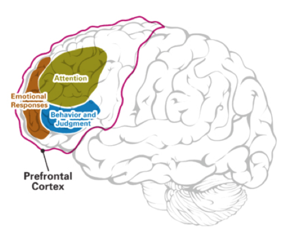
Image 1.5 is a diagrammatic representation of the prefrontal cortex and its functions. Prefrontal cortex plays an important role in attentional processes and is thought to be involved in the pathophysiology of ADHD. Image source - The Executive Functions of the Frontal Lobes [online image]. (2014) Human Physiology Academy. http://humanphysiology.academy/Neurosciences%202015/Chapter%203/P.3.1%20Cortical%20Functions.html
Understanding ADHD brain’s physiology through Neuroimaging-
Magnetic resonance imaging (MRI) cannot be used to diagnose ADHD but it has been performed by researchers to compare the anatomical differences between ADHD and normal children's brains. Specific regions of focus have included the cerebellum, corpus callosum, basal ganglia, and prefrontal regions in addition to assessing variations in overall brain volume. Grey and white matter variations have also been studied.
In normal brains, the cortex, which is the top layer and is involved in memory, attention, and language, is shown to thicken before the age of 11-13, but MRI scans of ADHD brains have revealed slower development, notably in frontal and temporal lobe regions of the cortex. ADHD brains are also observed to have smaller basal ganglia. These parts work together rather than independently to build networks that control functions such as attention, movement, language, etc. The functioning of these networks varies to allow the execution of different tasks.
For instance, while we are listening and paying attention in a classroom when the teacher is giving a lecture, the functioning of these networks involved in processing information will normally increase, but the activity of networks engaged in daydreaming would typically decrease. These networks' activity in the brain are discovered to be disrupted in ADHD. Some brain networks aren't turned on enough, while others are turned on too much when children with ADHD perform any particular task.
Comments
Post a Comment